
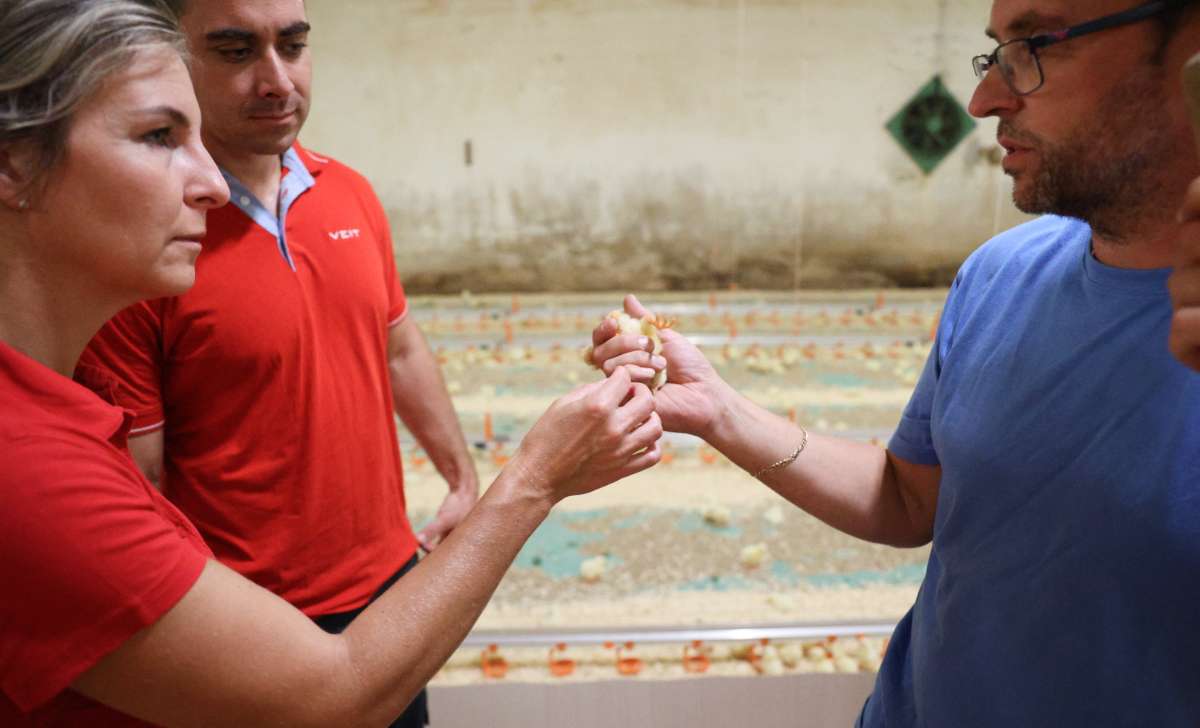
La coccidiosis en pollos es una de las enfermedades más perjudiciales desde el punto de vista económico en la producción avícola comercial. Está causada por protozoos del género Eimeria y afecta prácticamente a todas las parvadas en algún momento de su ciclo productivo. Según las estimaciones, su coste global asciende a aproximadamente 10,4 mil millones de libras esterlinas al año (Blake, 2025).
¿Qué es la coccidiosis en pollos?
La coccidiosis es una enfermedad intestinal causada por protozoos del género Eimeria. Existen siete especies capaces de infectar a los pollos, cada una de ellas localizada en diferentes segmentos del tracto digestivo.
Eimeria tenella coloniza los ciegos y provoca graves lesiones hemorrágicas. Por su parte, E. acervulina, E. maxima y E. brunetti afectan la absorción de nutrientes, a menudo sin presencia visible de sangre (Dalloul y Lillehoj, 2006).
Los ooquistes esporulan en la cama cálida y húmeda en un plazo de 24 a 48 horas. Las altas densidades de alojamiento favorecen la transmisión fecal-oral y aceleran la propagación de la enfermedad.
Signos y síntomas
Los signos más comunes incluyen letargo, agrupamiento de las aves, plumaje erizado, crestas pálidas, reducción del consumo de alimento y pérdida repentina de peso. En los brotes graves, la mortalidad puede aumentar rápidamente, en ocasiones en cuestión de pocos días (Ahmad et al., 2024).
La enfermedad altera la digestión y reduce el rendimiento productivo. Por esta razón, el peso corporal suele ser la primera señal medible de infección, especialmente porque los síntomas físicos iniciales suelen pasar desapercibidos.
Las parvadas monitorizadas mediante sistemas automáticos de pesaje diario suelen mostrar descensos característicos en el peso entre dos y tres días antes de que aparezcan los síntomas visibles, lo que permite tomar decisiones terapéuticas de forma más temprana.
Lo que pueden revelar las heces
La apariencia de las heces es un indicador diagnóstico fiable. Las deposiciones sanguinolentas de color marrón oscuro procedentes de los ciegos son características de una infección por E. tenella. La presencia de heces anaranjadas, mucosas o acuosas, por su parte, suele indicar una infección por E. maxima o E. brunetti.
Las infecciones subclínicas, en cambio, a menudo se manifiestan únicamente mediante heces más blandas y un deterioro progresivo de la calidad de la cama (Dalloul y Lillehoj, 2006).
Opciones de tratamiento
En la producción comercial, los coccidiostáticos incorporados al alimento constituyen la principal línea de defensa preventiva. Una vez que se establece un brote clínico, el daño intestinal se acumula rápidamente y resulta difícil de revertir (Ahmad et al., 2024). Por ello, la prevención mediante suplementación continua en la dieta suele ser más eficaz que el tratamiento curativo.
Cuando la infección clínica se confirma mediante el recuento de ooquistes en heces o mediante examen post mortem, las opciones terapéuticas autorizadas incluyen amprolio en el agua de bebida durante cinco a siete días, sulfonamidas activas frente a múltiples especies durante tres a cinco días y toltrazuril, que actúa sobre todas las fases del ciclo de vida del parásito en un único tratamiento (Ahmad et al., 2024).
Las vitaminas K y A favorecen la recuperación intestinal después de la infección. Antes de elegir cualquier protocolo terapéutico, se recomienda consultar con un veterinario.
Métodos naturales de prevención
Los enfoques naturales se centran principalmente en la prevención. Diversos estudios han demostrado que los compuestos derivados del orégano, los ácidos orgánicos y determinados probióticos pueden reducir la excreción de ooquistes, aunque no sustituyen a los medicamentos autorizados durante los brotes activos (Ahmad et al., 2024).
Las vacunas vivas basadas en ooquistes, utilizadas en sistemas de ponedoras y reproductoras, promueven una inmunidad controlada y reducen la dependencia de los coccidiostáticos incorporados al alimento (Cobb-Vantress, 2021).
Prevención y detección temprana mediante el monitoreo del peso
Una estrategia eficaz de prevención combina una adecuada gestión de la cama, el uso de coccidiostáticos en la alimentación, la vacunación y el monitoreo del peso corporal. El control de la humedad de la cama es esencial, ya que las condiciones húmedas favorecen la esporulación de los ooquistes. Asimismo, la rotación de anticoccidianos ayuda a reducir el riesgo de resistencia a los medicamentos.
La báscula automática para aves BAT2 Connect registra de forma continua el peso individual de las aves sin alterar el comportamiento de la parvada, permitiendo detectar desviaciones en el crecimiento antes de que aparezcan los síntomas clínicos.
El pesaje manual mediante la báscula avícola BAT1 añade una capa adicional de evaluación del bienestar animal. La manipulación individual permite valorar la condición corporal, el estado de hidratación y el llenado del tracto digestivo junto con el registro del peso.
Las tendencias de peso entre diferentes sesiones y naves pueden analizarse mediante BAT Cloud. Esto permite identificar si los problemas recurrentes de crecimiento se concentran en una nave específica, lo que a menudo puede indicar problemas estructurales, como grietas en el suelo o espacios donde los ooquistes sobreviven entre lotes de aves.
Los registros de captura y pesaje documentan el rendimiento productivo de la parvada antes, durante y después de un brote de coccidiosis.
Referencias citadas
1.) Ahmad, R., Yu, Y.-H., Hua, K.-F., Chen, W.-J., Zaborski, D., Dybus, A., Hsiao, F.S.-H., and Cheng, Y.-H. (2024). Management and control of coccidiosis in poultry: a review. Animal Bioscience, 37(1), 1–15. https://doi.org/10.5713/ab.23.0189
2.) Blake, D.P. (2025). Eimeria of chickens: the changing face of an old foe. Avian Pathology, 54(3), 267–278. https://doi.org/10.1080/03079457.2024.2441180
3.) Cobb-Vantress (2021). Cobb Breeder Management Guide. Cobb-Vantress Inc. https://www.cobbgenetics.com/assets/Cobb-Files/Breeder-Management-Guide.pdf
4.) Dalloul, R.A. and Lillehoj, H.S. (2006). Poultry coccidiosis: recent advancements in control measures and vaccine development. Expert Review of Vaccines, 5(1), 143–163. https://doi.org/10.1586/14760584.5.1.143